


Ceramic Partial Restorations (Anterior and Posterior Ceramic Restorations upto 6 units)
Restorative Ceramic Smile Makeover
*The requirements for participation are the same for both subcategories.
Ceramic restorations
Requirements
This category includes restorative treatments with ceramic materials. The restorations could be:
1) Partial (Anterior and/or Posterior Ceramic Restorartions upto 6 units)
2) Restorative Ceramic Smile Makeover
Judging criteria
The jury anonymously evaluates the submissions in the category according to the following criteria:
Diagnosis and treatment plan
Preparation
Restorations
Minimal invasiveness
Photo documentation
Occlusion
General treatment execution
Case description
Submissions must include a short description of the clinical case in English, not longer than 6,000 symbols.
Descriptions must outline:
Diagnosis
Treatment plan
Materials that were used according to the treatment protocol
Short summary of the clinical case
General photo documentation guidelines
1. Full facial front, wide smile
• 1:10 magnification.
• Patient’s smile is wide and natural.
• Whole face is visible, the top of the hair and the bottom of the neck can be cropped out.
• Both dental arches are visible.
• Background shadows must be reduced.
• The philtrum and nose are centrally positioned in the frame.
• Patient’s head is straight up.
• Camera is levelled with the nose.
• Camera is oriented on the interpupillary line and the vertical midline of the face.
• Focus is on the teeth.
• Background is plain and undistracting.

2. Frontal smile, non-retracted
• 1:2 magnification.
• Lips and teeth are visible, natural smile.
• Camera is levelled with the smile.
• Camera is oriented on the incisal plane and the midline, as long as they coincide with the interpupillary line and the vertical midline of the face.
• In case of asymmetry, it must be apparent in the image.
• Focus is on the central incisors, the laterals, and the canines.
• Image is cropped to the mouth edges and a little above the upper lip.

3. Full smile, left and right, non-retracted (frontal sagittal view)
• 1:2 magnification.
• Lips and teeth are visible, wide smile.
• Image is taken from the side so that the lateral incisor is in the center of the frame.
• The contralateral incisor and the canine must be visible, depending on arch size.
• The vertical midline is the lateral.
• Focus is on the lateral.
• Background might be necessary.
• The nose, the top of the cheeks and the chin are cropped out of the image.

4. Frontal view, retracted (central view, teeth apart)
• 1:2 magnification.
• Upper and lower teeth are slightly parted so that the incisal edges are visible.
• Show as much of the gingiva as possible.
• No lips and retractors visible in the frame.
• Use the midline of the face as the vertical midline of the image.
• The incisal plane of the upper arch is the horizontal midline of the image.
• If there is asymmetry, it must be apparent in the picture.
• Camera is levelled with the teeth.
• Focus is on the central incisors, the laterals, and the canines.

5. Frontal view, upper and lower teeth, bite
• 1:1 magnification.
• The midline of the incisors is the vertical midline of the image.
• The horizontal midline of the image bisects the upper incisors.
• Selected 4-6 teeth are cropped out of the image.
• Show as much of the gingiva as possible.
6. Frontal view of treated teeth, teeth apart (upper or lower: show teeth that have been treated)
• 1:1 magnification.
• The upper and lower incisors are visible (the ones that have been treated), the gingiva adjacent to the teeth is also visible. The retracted lip and antagonists must not be visible. At this level of magnification, 4-6 upper teeth or 6-8 lower teeth are visible.
• Camera is levelled with the teeth.
• The central incisors are the midline of the image.
Upper front, teeth apart
• No retractors are visible.
• Show as much of the gingiva as possible.
• The philtrum and the midline of the incisors are the vertical midline of the image.
• No antagonists are visible.
• Selected 4-6 teeth are cropped out of the image.
Lower front, teeth apart
• The midline of the incisors is the vertical midline of the image.
• Show as much of the gingiva as possible.
• No antagonists are visible.
• Selected 4-6 teeth are cropped out of the image.

Removed saliva, plaque, food, crown-cement residue.
Use make-up when photographing the patient’s face.
The camera is centered at an appropriate angle.
Use a backdrop when necessary.
Retractors, mirrors and other elements must not be in the field of view.
Images must have good contrast, brightness and white balance.
Photos must be symmetric and cropped properly.
Objects in photos must be scaled appropriately.
Exclude any personal information, names and initials patient’s and dental professional’s).
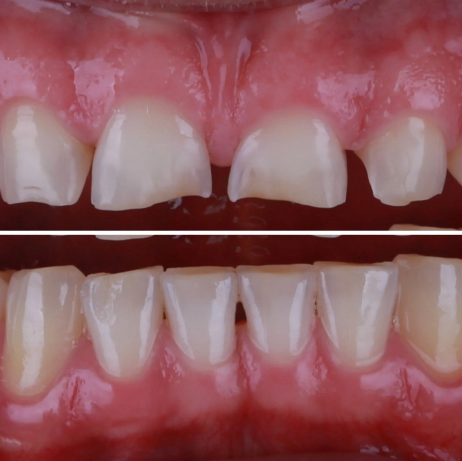
Initial photo documentation (before treatment)

7. Recommended: upper and lower front using a contraster.
Upper front with contraster
• 1:2 magnification.
• Use a contrasting device.
• Color may be other than black.
• No shadows on the contraster.
Lower front with contraster
• 1:2 magnification.
• Use a contrasting device.
• Color may be other than black.
• No shadows on the contraster.
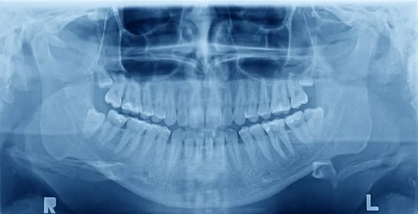
8. Please, include X- ray before obtuariation.
During treatment
Clinical stage:
1. Prepped teeth.
2. Temporary restorations.
Laboratory stage:
1. Dental models.
2. Recommended: finished restoration on the model.
After treatment
Same photo documentation as in Before Treatment.
In cases of direct restorations, include:
Cavity preparation before obturation.
At least 1 photo of the composite build up.
Important! X-ray after obturation.
When restoring incisal edges – protrusion and/or laterotrusion (when canines were restored as well).